
Science has utilised light for centuries. It underpins our attempts to understand it – the speed of light is one of the primary scientific constants, and light allows us to detect everything from the gravitational pull of far-away galaxies and to manipulate the intricacies of our own DNA. Kishan Dholakia specialises in the science of light, particularly as it relates to biological discovery and healthcare. He leads the Optical Manipulation Group within the Centre for Biophotonics in the School of Physics and Astronomy, which has an extensive track record in the development of using shaped light beams for imaging studies.
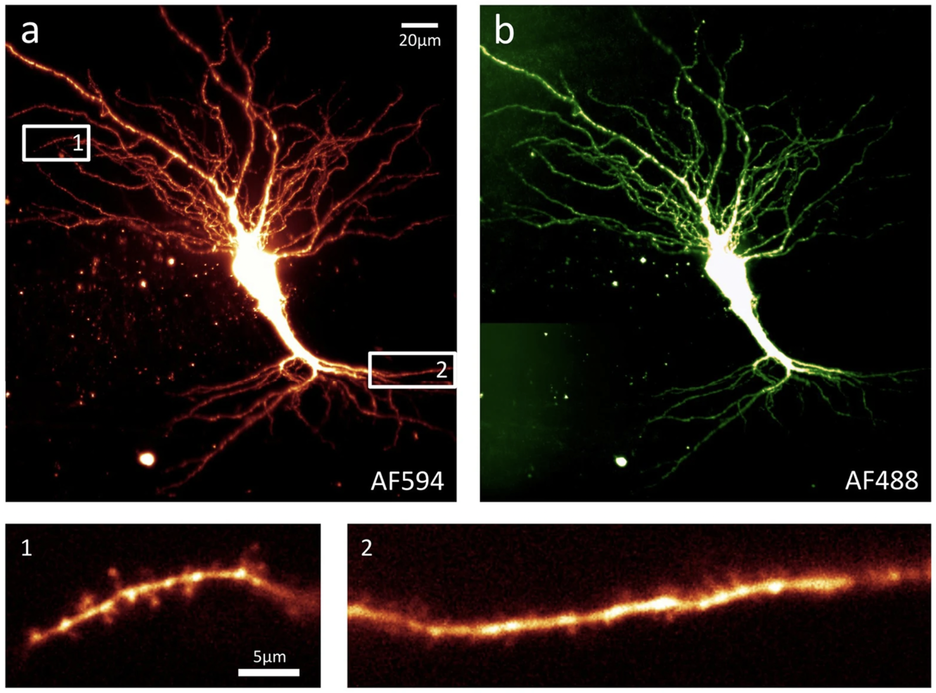
Optical imaging for biological applications is undergoing a revolution, reaching ever higher resolutions and increasing its capture area to image increasingly large structures. This is fuelled in part by light sheet fluorescence microscopy (LSFM), which irradiates a section of the subject with a thin ‘sheet’ of light and the fluorescence or scattered light is collected at right angles to the illumination. The method is very appealing when compared to standard microscopes, which illuminate and collect light through the same optics, as it allows rapid 3D imaging of live specimens and offers high contrast due to the targeted illumination. LSFM also benefits from very low toxicity due to dramatically reduced light exposure: 10 to 1,000 times lower than conventional imaging, as only the part of the sample from which one wishes to extract a signal is irradiated. LSFM is the tool of choice for neuroscience and developmental biology. It also has great potential for drug discovery, histopathology, and phenotypical studies as it allows rapid (sub-second) imaging of tens to hundreds of samples.
The LSFM methodology has had three challenges that, until recently, have limited its applicability. Firstly, scientists have struggled to create a thin, sheet-like beam of light that extends right across the field of view of the sample to provide uniform image quality across the whole field of vision. This difficulty is compounded by the need to keep the illumination level as low as possible to avoid any damage to samples from excessive light exposure. Finally, there is the ostensibly contradictory need to achieve increased light penetration into biological samples to allow imaging at more than just the surface level. Until now, these three challenges have restricted biomedical understanding and the ability to achieve high sensitivity and specificity in early diagnosis. However, the Optical Research Group’s approach of using the Airy beam, a transverse light pattern in two dimensions that does not change as the beam moves across the sample, addresses all three of these drawbacks. As a result, the technology can improve imaging of many biological systems, including the whole mouse brain, samples from multiple types of cancer, and model organisms such as zebra fish.
Prof. Dholakia’s group showed that they could overcome the restrictions in LSFM using an airy beam in 2014. The airy beam has a unique structural property that allows the retrieval of high-resolution information via a straightforward mathematical algorithm to create an image of the whole sample. The team showed that microscope utilising airy beams could be developed into a compact solution to imaging in neuroscience in 2016, and that the approach is also compatible with more advanced fluorescence techniques in 2017. In 2018, the group’s work tailoring the Airy beam to overcome degradation issues allowed it to achieve deeper penetration into samples.
The University’s Airy beam LSFM technology was exclusively licensed to M Squared Lasers Ltd, a Scottish company headquartered in Glasgow and an international leader in photonics technology. In 2016 a new subsidiary, M Squared Life Ltd in 2016, was established specifically to develop and sell the “Aurora” light sheet microscope, a new scientific instrument based on the Airy beam developed at St Andrews. This has created several jobs in Scotland and secured significant investment to grow both businesses.
Aurora has since been used to advance our understanding of diseases and processes in neuroscience and developmental biology in Universities, Research Institutes and National Laboratories across the world. Aurora units can be found across the UK, Europe, South Africa, Australia, and the USA. Compared with competing technologies, Aurora enables end users to image a wider variety of biological samples with less photon damage, and to do so more rapidly and with unprecedented image quality over a larger volume. These benefits are enabling new imaging approaches not previously possible, particularly for live samples.